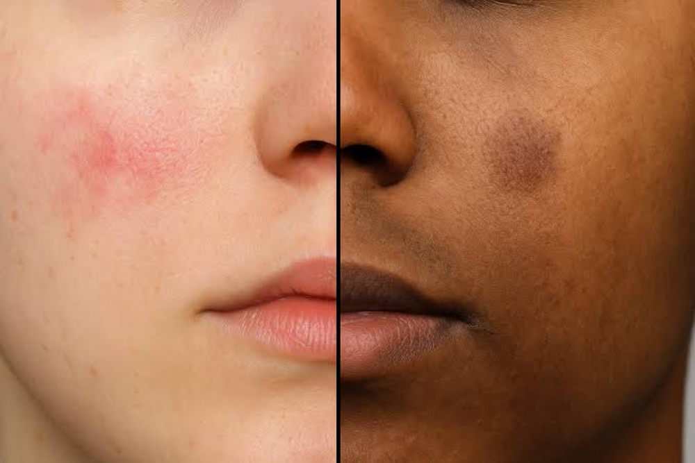
You’ve finally cleared an active pimple, but in its place sits a stubborn spot that lingers for months. Many people lump all post-breakout marks into one category, but biologically, they fall into two completely different types: Post-Inflammatory Erythema (PIE) and Post-Inflammatory Hyperpigmentation (PIH).
Treating a red mark like a brown spot won’t yield results—which is why identifying the underlying cause is key.
| Feature | Post-Inflammatory Erythema (PIE) | Post-Inflammatory Hyperpigmentation (PIH) |
| Color Profile | Pink, red, or purplish flat spots | Light brown, dark brown, or black flat spots |
| Root Biological Cause | Damaged, dilated micro-capillaries (blood vessels) | Over-stimulated melanocytes (pigment cells) |
| Who Gets It Most? | Fitzpatrick Skin Types I–III (fairer skin tones) | Fitzpatrick Skin Types IV–VI (deeper skin tones) |
| The “Blanch Test” | Turns white briefly when pressed with a glass slide | Stays the same color when pressed |
| Best Target Treatments | Vascular Lasers, Niacinamide, Centella, LED | Tyrosinase Inhibitors, Chemical Peels, CO2 Laser |
Why the Difference Dictates Your Treatment
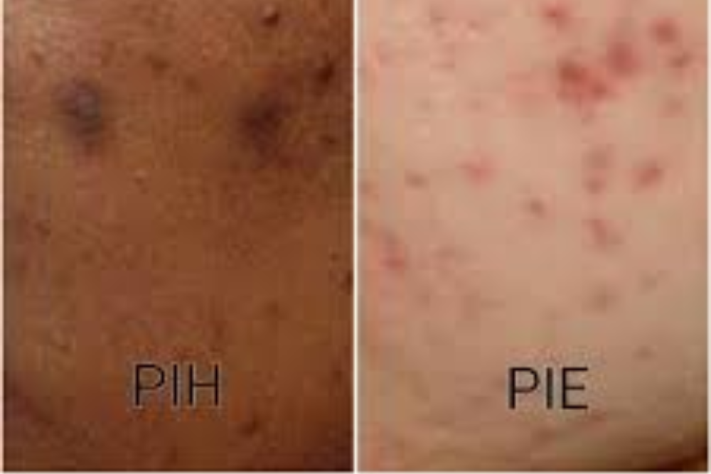
- If You Have PIE (Red Marks):Because PIE is a vascular issue (damaged capillaries near the surface), standard pigment-fading ingredients like Hydroquinone won’t help. Focus on vascular-calming ingredients (Niacinamide, Azelaic Acid) and vascular light therapies that shrink expanded blood vessels.
- If You Have PIH (Brown Marks):Because PIH is caused by excess melanin, treatment requires pigment-blocking agents (Tranexamic Acid, Kojic Acid, Vitamin C) combined with gentle chemical exfoliants or modified fractional lasers that lift excess melanin out of the epidermis.